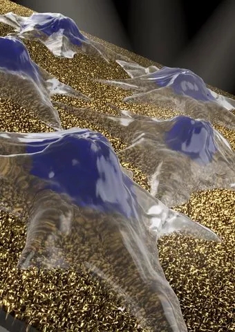
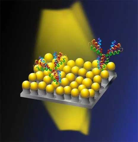
Release date: 2018-03-05 Researchers from the Okinawa Institute of Science and Technology (OIST) have invented a plasmonic nanosensor that monitors cell proliferation in real time and has other potential applications. The study was published in the recent issue of ACS Applied Materials and Interfaces. Revealing the cell proliferation process is an important insight into the health and function of cells and tissues. The figure shows the cell (blue mountain shape) on top of the nano mushroom-like structure. This "nano mushroom" contains a silica stem and a gold cap. This sensor is expected to monitor cell proliferation in real time. Image source: OIST The most attractive thing about this material is that it allows cells to survive for a long time. “Usually, when you put living cells on nanomaterials, because the quality of nanomaterials is toxic, it kills cells,†said Dr. Nikhil Bhalla, the post-doctoral researcher of the paper. “However, using our The material, the cells survived for more than 7 days. The nanoparticle material is also highly sensitive: it can detect micro-proliferation of 16 of 1000 cells." These materials look like ordinary glass pieces. However, the surface is covered with tiny nano-granular structures called nano-mushrooms, with silica stems and gold hats. These constituent biosensors are capable of detecting molecular level interactions. The biosensor works by using nano-mesh as an optical antenna. When white light passes through the thin slices of nanoparticles, the nano-muse absorbs and scatters some of the light, changing its properties. The absorbance and scattering of light is determined by the size, shape and material of the nanomaterial, and more importantly, it is also affected by any medium close to the nano mushroom, such as cells placed on the nanoflakes. By measuring changes in the appearance of light on the other side of the sheet, researchers can detect and monitor processes that occur on the sensor surface, such as cell division. “Normally, you have to add some labels, such as dyes or molecules, to your cells to calculate the number of cell divisions,†Dr. Bhalla said. “However, with our approach, nanobots can sense them directly.†Although nanomaterials have great advantages for detection, the production of large-scale nanomaterials is challenging because it is difficult to ensure uniformity across the surface of the material. For this reason, biosensors for routine clinical examination, such as disease testing, are still lacking. To cope with this problem, researchers have developed a new type of printing technology to make large-scale nano mushroom biosensors. Through their method, they were able to develop a 2.5 cm silica substrate consisting of approximately 1 million mushroom-like structures. “Our technology is like printing a stamp, covering it with biomolecule ink and then printing it on a thin piece of nanoparticles,†said Dr. Shivani Sathish, co-author of the paper. Biomolecules increase the sensitivity of a material, which means it can perceive very low concentrations of substances, such as antibodies, making it possible to detect disease early. Using their innovative printing technology, OIST researchers have developed a nanoplasma material that contains millions of biomolecules resembling mushroom-like structures. Image source: OIST “Using our approach, we have the potential to create a highly sensitive biosensor that can even detect a single molecule,†said Dr. Bhalla, the lead author of the paper. From electronics to food production to medicine, plasma and nanoparticle sensors provide an important tool in many areas. Reference materials: [1] Nanomushroom Sensors: One Material, Many Applications Source: Health New Vision Tcm Extract for traditonal medicine,Traditopnal Medicine For Sheep,Largehead Atractylodes Rhizome Extract powder Sichuan Aibang Weiye Biological Engineering Co., Ltd. , https://www.aibangpharm.com
