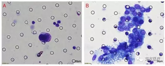
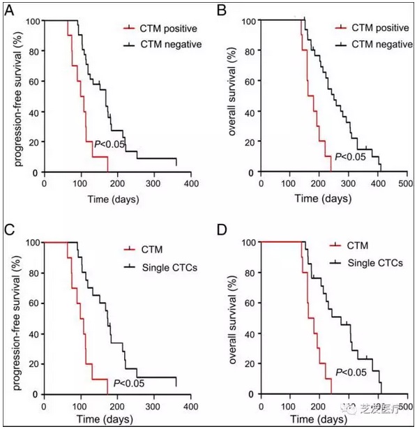
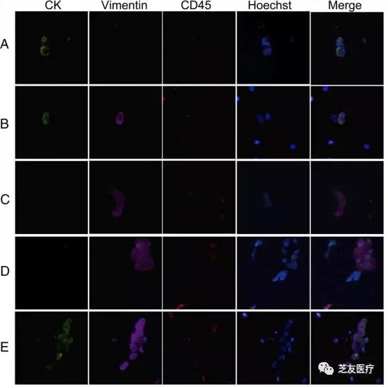
The latest scientific research cooperation between Wuhan Union Hospital and Zhiyou Medical: Circulating tumor micro-embolization has guiding significance for the prognosis of patients with gastric cancer Lead: Gastric cancer (GC) is one of the most common malignant tumors in China. Professor Zhang Tao from the Cancer Center of Wuhan Union Hospital, through the CTCBIOPSY® platform, is used to separate circulating tumor cells (CTCs) and circulating tumor microtubules in peripheral blood of patients with gastric cancer. (Circulating tumor microemboli, CTM), to explore the relationship between CTCs/CTM detection and gastric cancer staging and other clinical indicators, CTM detection and prognosis of patients with stage IV gastric cancer. Background introduction: Gastric cancer is a high-risk cancer in Southeast Asia and one of the most common cancers in China. It has a high morbidity and mortality rate and has a tendency to become younger in recent years. As a very promising liquid biopsy index, CTCs are widely studied in gastric cancer. CTM refers to circulating tumor microtubules formed by aggregation of three or more CTCs, which has stronger invasive and distant metastatic ability. Because traditional density gradient centrifugation and immunomagnetic bead capture methods are difficult to capture CTM, CTM is currently rarely studied in gastric cancer. CTCBIOPSY ® is an automated CTCs/CTM separation and capture device based on patented filter micro-screen technology independently developed by Zhiyou Medical. It has strong capture ability for CTCs and CTM. The device uses Diff dye to rapidly stain cells and perform morphological identification. . Researchers from Union Hospital used CTCBIOPSY ® to isolate CTCs and CTM from peripheral blood of patients with gastric cancer, and identified their typing characteristics using downstream immunofluorescence techniques. The marker was the epithelial marker Cytokeratin (CK). The interstitial marker vimentin (Vimentin), the white blood cell common antigen CD45. The researchers then explored the relationship between CTCs and CTM detection and clinical indicators, and did an in-depth analysis of whether CTM was detected in patients. research content: In this study, 30 healthy individuals and 86 gastric cancer patients were enrolled, 5 of whom were stage I, 15 were stage II, 25 were stage III, and 41 were stage IV. 5 mL of peripheral blood samples were collected prior to systemic treatment. CTCBIOPSY ® was used to separate CTCs and CTM, and these CTCs and CTM were identified by immunofluorescence, which were divided into epithelial type (CK+/Vimentin-/CD45-), epithelial interstitial type (CK+/Vimentin+/CD45-) and interstitial Type (CK-/Vimentin+/CD45-). Among 30 healthy people, there were no CTCs and CTM. Of the 86 patients with gastric cancer, 51 had CTCs with a mean value of 1.81. In patients with stage I, II, III and IV, the mean values ​​of CTCs were 1.40, 0.67, 1.24, and 2.71 (Fig. 1A); CTM was detected in 3 of 33 patients in stage I to IIIB with a mean of 0.12 (0-2), and 13 of 53 patients with stage IIIC to IV detected CTM. The mean is 1.26 (0-22) (Figure 1B). Among the various clinical indicators of patients, CTM detection was significantly associated with clinical stage (P=0.05). The PFS and OS of CTM positive patients were significantly lower than those of CTM negative patients (P<0.05) (Fig. 2A, B). The PFS and OS of CTM positive patients were significantly lower than those with only one CTCs positively detected (P). <0.05) (Fig. 2C, D), CTM was an independent prognostic factor for poorer PFS and OS in stage IV patients (PFS, P = 0.016; OS, P = 0.003). Immunofluorescence experiments showed that there were three phenotypes of epithelial (Fig. 3A), epithelial interstitial (Fig. 3B) and interstitial (Fig. 3C) in CTCs, and interstitial type of CTM (Fig. 3D) and epithelial interstitial Two phenotypes (Figure 3E). ▲ Figure 1 CTCs and CTM (Diff Rapid Staining) detected in patients with gastric cancer ▲ Figure 2 CTCs/CTM detection and patient PFS and OS relationship ▲ Figure 3 Immunofluorescence staining of CTCs and CTM Analysis conclusion: By CTCBIOPSY ® platform for the detection of gastric cancer patients CTCs and CTM, compatible downstream immunofluorescence techniques on these CTCs and CTM were phenotypically characterized and found independent prognostic CTM in Ⅳ patients are poorer PFS and OS factor. With its good sensitivity and specificity, CTCBIOPSY ® can effectively detect CTCs and CTM in peripheral blood of patients, help clinicians evaluate the condition of cancer patients, and develop personalized treatment plans. This article is the latest collaborative research results of the team of Professor Zhang Tao from the Cancer Center of Wuhan Union Hospital and Zhiyou Medical! Professor Zhang Tao personal and team profile: Zhang Tao, MD, professor, chief physician, doctoral tutor. Deputy Director of the Cancer Center of the Union Hospital of Tongji Medical College, Huazhong University of Science and Technology, deputy director of the Department of Oncology. Professor Zhang Tao's research group has been committed to research on cancer stem cells and tumor biotherapy for many years. In recent years, he has undertaken a total of 7 national natural science funds and provincial and ministerial level projects. His research results have published more than 20 SCI papers, respectively. "Blood", "Cancer Research", "Journal of Immunotherapy" and other magazines provide a theoretical basis and practical basis for targeted therapy, enhanced efficacy and biological treatment of malignant tumors. This article is the original translation of Zhiyou Medical. Original source: Zheng, X., et al. (2017). "Detection of Circulating Tumor Cells and Circulating Tumor Microemboli in Gastric Cancer." Transl Oncol 10(3): 431-441. Inverted Fluorescent Microscope Inverted Cell Researching Microscope,High-Level Clinic Inverted Researching Microscope,High-Level Medical Inverted Researching Microscope,Medical Inverted Researching Microscope Ningbo ProWay Optics & Electronics Co., Ltd. , https://www.proway-microtech.com